

What Method Is Used By Animals For Growth And Repair Of Their Tissues?

In biology, regeneration is the process of renewal, restoration, and tissue growth that makes genomes, cells, organisms, and ecosystems resilient to natural fluctuations or events that cause disturbance or impairment.[i] Every species is capable of regeneration, from bacteria to humans.[2] [iii] Regeneration can either be complete[4] where the new tissue is the same as the lost tissue,[four] or incomplete[5] where after the necrotic tissue comes fibrosis.[five]
At its most unproblematic level, regeneration is mediated by the molecular processes of cistron regulation and involves the cellular processes of cell proliferation, morphogenesis and jail cell differentiation.[6] [7] Regeneration in biological science, however, mainly refers to the morphogenic processes that characterize the phenotypic plasticity of traits allowing multi-cellular organisms to repair and maintain the integrity of their physiological and morphological states. In a higher place the genetic level, regeneration is fundamentally regulated by asexual cellular processes.[eight] Regeneration is different from reproduction. For instance, hydra perform regeneration but reproduce by the method of budding.
The hydra and the planarian flatworm accept long served every bit model organisms for their highly adaptive regenerative capabilities.[ix] Once wounded, their cells become activated and restore the organs back to their pre-existing state.[ten] The Caudata ("urodeles"; salamanders and newts), an society of tailed amphibians, is possibly the most adept vertebrate group at regeneration, given their capability of regenerating limbs, tails, jaws, eyes and a variety of internal structures.[ii] The regeneration of organs is a common and widespread adaptive capability amid metazoan creatures.[9] In a related context, some animals are able to reproduce asexually through fragmentation, budding, or fission.[8] A planarian parent, for example, will constrict, split in the middle, and each half generates a new end to form two clones of the original.[11]
Echinoderms (such as the sea star), crayfish, many reptiles, and amphibians exhibit remarkable examples of tissue regeneration. The case of autotomy, for example, serves equally a defensive office equally the animal detaches a limb or tail to avoid capture. Afterwards the limb or tail has been autotomized, cells move into action and the tissues will regenerate.[12] [xiii] [14] In some cases a shed limb tin itself regenerate a new private.[15] Limited regeneration of limbs occurs in most fishes and salamanders, and tail regeneration takes place in larval frogs and toads (but not adults). The whole limb of a salamander or a triton will grow again and again after amputation. In reptiles, chelonians, crocodilians and snakes are unable to regenerate lost parts, merely many (not all) kinds of lizards, geckos and iguanas possess regeneration chapters in a high degree. Usually, information technology involves dropping a section of their tail and regenerating it equally part of a defense force machinery. While escaping a predator, if the predator catches the tail, it volition disconnect.[16]
Ecosystems [edit]
Ecosystems can be regenerative. Following a disturbance, such equally a fire or pest outbreak in a forest, pioneering species will occupy, compete for space, and establish themselves in the newly opened habitat. The new growth of seedlings and community assembly process is known as regeneration in ecology.[17] [xviii]
Cellular molecular fundamentals [edit]
Blueprint formation in the morphogenesis of an animate being is regulated by genetic induction factors that put cells to work after damage has occurred. Neural cells, for example, express growth-associated proteins, such as GAP-43, tubulin, actin, an array of novel neuropeptides, and cytokines that induce a cellular physiological response to regenerate from the damage.[19] Many of the genes that are involved in the original development of tissues are reinitialized during the regenerative process. Cells in the primordia of zebrafish fins, for example, express four genes from the homeobox msx family during development and regeneration.[20]
Tissues [edit]
"Strategies include the rearrangement of pre-existing tissue, the use of adult somatic stem cells and the dedifferentiation and/or transdifferentiation of cells, and more one mode can operate in different tissues of the same animal.[ane] All these strategies outcome in the re-establishment of appropriate tissue polarity, structure and grade."[21] : 873 During the developmental process, genes are activated that serve to modify the properties of cell as they differentiate into different tissues. Development and regeneration involves the coordination and organization of populations cells into a blastema, which is "a mound of stem cells from which regeneration begins".[22] Dedifferentiation of cells means that they lose their tissue-specific characteristics as tissues remodel during the regeneration procedure. This should non exist dislocated with the transdifferentiation of cells which is when they lose their tissue-specific characteristics during the regeneration process, and and then re-differentiate to a different kind of cell.[21]
In animals [edit]
Arthropods [edit]
Arthropods are known to regenerate appendages following loss or autotomy.[23] Regeneration among arthropods is restricted by molting such that hemimetabolous insects are capable of regeneration only until their concluding molt whereas about crustaceans tin regenerate throughout their lifetimes.[24] Molting cycles are hormonally regulated in arthropods,[25] although premature molting tin can be induced by autotomy.[23] Mechanisms underlying appendage regeneration in hemimetabolous insects and crustaceans are highly conserved.[26] During limb regeneration species in both taxa class a blastema[27] following autotomy with regeneration of the excised limb occurring during proecdysis.[25] Limb regeneration is also present in insects that undergo metamorphosis, such as beetles, although the cost of said regeneration is a delayed pupal stage.[28] Arachnids, including scorpions, are known to regenerate their venom, although the content of the regenerated venom is different from the original venom during its regeneration, as the venom book is replaced before the active proteins are all replenished.[29]
Annelids [edit]
Many annelids (segmented worms) are capable of regeneration.[thirty] For example, Chaetopterus variopedatus and Branchiomma nigromaculata can regenerate both anterior and posterior body parts afterward latitudinal bisection.[31] The relationship between somatic and germline stem cell regeneration has been studied at the molecular level in the annelid Capitella teleta.[32] Leeches, however, appear incapable of segmental regeneration.[33] Furthermore, their close relatives, the branchiobdellids, are also incapable of segmental regeneration.[33] [30] However, sure individuals, like the lumbriculids, can regenerate from but a few segments.[33] Segmental regeneration in these animals is epimorphic and occurs through blastema germination.[33] Segmental regeneration has been gained and lost during annelid development, as seen in oligochaetes, where head regeneration has been lost three separate times.[33]
Along with epimorphosis, some polychaetes similar Sabella pavonina experience morphallactic regeneration.[33] [34] Morphallaxis involves the de-differentiation, transformation, and re-differentation of cells to regenerate tissues. How prominent morphallactic regeneration is in oligochaetes is currently not well understood. Although relatively under-reported, it is possible that morphallaxis is a common mode of inter-segment regeneration in annelids. Following regeneration in L. variegatus, past posterior segments sometimes become inductive in the new body orientation, consistent with morphallaxis.
Following amputation, most annelids are capable of sealing their torso via rapid muscular contraction. Constriction of body muscle tin lead to infection prevention. In sure species, such as Limnodrilus, autolysis can be seen within hours after amputation in the ectoderm and mesoderm. Amputation is also idea to crusade a large migration of cells to the injury site, and these course a wound plug.
Echinoderms [edit]
Tissue regeneration is widespread among echinoderms and has been well documented in starfish (Asteroidea), sea cucumbers (Holothuroidea), and sea urchins (Echinoidea). Bagginess regeneration in echinoderms has been studied since at to the lowest degree the 19th century.[35] In improver to appendages, some species can regenerate internal organs and parts of their central nervous system.[36] In response to injury starfish can autotomize damaged appendages. Autotomy is the self-amputation of a body part, usually an bagginess. Depending on severity, starfish volition then go through a 4-week procedure where the appendage will be regenerated.[37] Some species must retain rima oris cells to regenerate an appendage, due to the demand for free energy.[38] The get-go organs to regenerate, in all species documented to engagement, are associated with the digestive tract. Thus, nearly knowledge nearly visceral regeneration in holothurians concerns this system.[39]
Planaria (Platyhelminthes) [edit]
Regeneration inquiry using Planarians began in the belatedly 1800s and was popularized by T.H. Morgan at the showtime of the 20th century.[38] Alejandro Sanchez-Alvarado and Philip Newmark transformed planarians into a model genetic organism in the beginning of the 20th century to study the molecular mechanisms underlying regeneration in these animals.[40] Planarians exhibit an boggling power to regenerate lost body parts. For example, a planarian split lengthwise or crosswise volition regenerate into ii divide individuals. In 1 experiment, T.H. Morgan found that a slice corresponding to ane/279th of a planarian[38] or a fragment with as few as x,000 cells tin can successfully regenerate into a new worm within one to ii weeks.[41] After amputation, stump cells form a blastema formed from neoblasts, pluripotent cells institute throughout the planarian trunk.[42] New tissue grows from neoblasts with neoblasts comprising between twenty and thirty% of all planarian cells.[41] Recent work has confirmed that neoblasts are totipotent since one single neoblast can regenerate an unabridged irradiated brute that has been rendered incapable of regeneration.[43] In lodge to foreclose starvation a planarian will use their own cells for free energy, this phenomenon is known equally de-growth.[ten]
Amphibians [edit]
Limb regeneration in the axolotl and newt has been extensively studied and researched. The nineteenth century studies of this subject are reviewed in Holland (2021).[44] Urodele amphibians, such as salamanders and newts, display the highest regenerative ability among tetrapods.[45] [44] Every bit such, they tin can fully regenerate their limbs, tail, jaws, and retina via epimorphic regeneration leading to functional replacement with new tissue.[46] Salamander limb regeneration occurs in ii principal steps. First, the local cells dedifferentiate at the wound site into progenitor to form a blastema.[47] Second, the blastemal cells will undergo cell proliferation, patterning, cell differentiation and tissue growth using similar genetic mechanisms that deployed during embryonic development.[48] Ultimately, blastemal cells will generate all the cells for the new construction.[45]

Axolotls tin can regenerate a variety of structures, including their limbs
After amputation, the epidermis migrates to cover the stump in ane–two hours, forming a structure called the wound epithelium (We).[49] Epidermal cells continue to migrate over the WE, resulting in a thickened, specialized signaling middle chosen the apical epithelial cap (AEC).[50] Over the adjacent several days there are changes in the underlying stump tissues that result in the formation of a blastema (a mass of dedifferentiated proliferating cells). As the blastema forms, pattern germination genes – such as HoxA and HoxD – are activated as they were when the limb was formed in the embryo.[51] [52] The positional identity of the distal tip of the limb (i.e. the autopod, which is the hand or human foot) is formed first in the blastema. Intermediate positional identities between the stump and the distal tip are and so filled in through a process called intercalation.[51] Motor neurons, muscle, and blood vessels abound with the regenerated limb, and reestablish the connections that were present prior to amputation. The fourth dimension that this entire procedure takes varies according to the age of the animal, ranging from about a month to around three months in the adult so the limb becomes fully functional. Researchers at Australian Regenerative Medicine Found at Monash University have published that when macrophages, which eat upwards material debris,[53] were removed, salamanders lost their ability to regenerate and formed scarred tissue instead.[54]
In spite of the historically few researchers studying limb regeneration, remarkable progress has been made recently in establishing the neotenous amphibian the axolotl (Ambystoma mexicanum) as a model genetic organism. This progress has been facilitated by advances in genomics, bioinformatics, and somatic cell transgenesis in other fields, that take created the opportunity to investigate the mechanisms of important biological backdrop, such as limb regeneration, in the axolotl.[48] The Ambystoma Genetic Stock Heart (AGSC) is a cocky-sustaining, convenance colony of the axolotl supported by the National Science Foundation as a Living Stock Collection. Located at the Academy of Kentucky, the AGSC is dedicated to supplying genetically well-characterized axolotl embryos, larvae, and adults to laboratories throughout the United States and abroad. An NIH-funded NCRR grant has led to the establishment of the Ambystoma EST database, the Salamander Genome Projection (SGP) that has led to the creation of the first amphibian gene map and several annotated molecular data bases, and the cosmos of the research community web portal.[55]
Frog model [edit]
Anurans (frogs) can merely regenerate their limbs during embryonic development.[56] Reactive oxygen species (ROS) appear to be required for a regeneration response in the anuran larvae.[57] ROS product is essential to activate the Wnt signaling pathway, which has been associated with regeneration in other systems.[57]
Once the limb skeleton has developed in frogs, regeneration does not occur (Xenopus can grow a cartilaginous spike later amputation).[56] The adult Xenopus laevis is used as a model organism for regenerative medicine. In 2022, a cocktail of drugs and hormones (1,4-DPCA, BDNF, growth hormone, resolvin D5, and retinoic acid), in a single dose lasting 24 hours, was shown to trigger long-term leg regenration in developed X. laevis. Instead of a unmarried spike, a paddle-shaped growth is obtained at the finish of the limb by xviii months.[58]
Hydra [edit]
Hydra is a genus of freshwater polyp in the phylum Cnidaria with highly proliferative stem cells that gives them the ability to regenerate their entire body.[59] Any fragment larger than a few hundred epithelial cells that is isolated from the body has the ability to regenerate into a smaller version of itself.[59] The high proportion of stem cells in the hydra supports its efficient regenerative power.[60]
Regeneration among hydra occurs as foot regeneration arising from the basal part of the body, and head regeneration, arising from the apical region.[59] Regeneration tissues that are cut from the gastric region comprise polarity, which allows them to distinguish between regenerating a head in the apical end and a foot in the basal terminate so that both regions are present in the newly regenerated organism.[59] Caput regeneration requires complex reconstruction of the surface area, while foot regeneration is much simpler, similar to tissue repair.[61] In both foot and head regeneration, however, in that location are ii distinct molecular cascades that occur once the tissue is wounded: early injury response and a subsequent, signal-driven pathway of the regenerating tissue that leads to cellular differentiation.[60] This early-injury response includes epithelial prison cell stretching for wound closure, the migration of interstitial progenitors towards the wound, jail cell death, phagocytosis of cell droppings, and reconstruction of the extracellular matrix.[lx]
Regeneration in hydra has been defined as morphallaxis, the procedure where regeneration results from remodeling of existing cloth without cellular proliferation.[62] [63] If a hydra is cut into ii pieces, the remaining severed sections grade 2 fully functional and independent hydra, approximately the aforementioned size as the two smaller severed sections.[59] This occurs through the exchange and rearrangement of soft tissues without the formation of new material.[60]
Aves (birds) [edit]
Owing to a limited literature on the bailiwick, birds are believed to have very limited regenerative abilities as adults. Some studies[64] on roosters accept suggested that birds can adequately regenerate some parts of the limbs and depending on the atmospheric condition in which regeneration takes identify, such equally age of the animal, the inter-human relationship of the injured tissue with other muscles, and the type of performance, can involve consummate regeneration of some musculoskeletal structure. Werber and Goldschmidt (1909) found that the goose and duck were capable of regenerating their beaks after partial amputation[64] and Sidorova (1962) observed liver regeneration via hypertrophy in roosters.[65] Birds are also capable of regenerating the hair cells in their cochlea following dissonance harm or ototoxic drug damage.[66] Despite this evidence, contemporary studies advise reparative regeneration in avian species is express to periods during embryonic evolution. An array of molecular biology techniques have been successful in manipulating cellular pathways known to contribute to spontaneous regeneration in chick embryos.[67] For instance, removing a portion of the elbow joint in a chick embryo via window excision or piece excision and comparing articulation tissue specific markers and cartilage markers showed that window excision allowed ten out of 20 limbs to regenerate and expressed joint genes similarly to a developing embryo. In dissimilarity, slice excision did not allow the joint to regenerate due to the fusion of the skeletal elements seen by an expression of cartilage markers.[68]
Similar to the physiological regeneration of pilus in mammals, birds tin can regenerate their feathers in guild to repair damaged feathers or to attract mates with their plume. Typically, seasonal changes that are associated with breeding seasons will prompt a hormonal bespeak for birds to begin regenerating feathers. This has been experimentally induced using thyroid hormones in the Rhode Island Red Fowls.[69]
Mammals [edit]
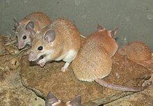
Spiny mice (Acomys cahirinus pictured hither) can regenerate pare, cartilage, nerves and musculus.
Mammals are capable of cellular and physiological regeneration, but accept more often than not poor reparative regenerative ability across the grouping.[1] [24] Examples of physiological regeneration in mammals include epithelial renewal (due east.g., skin and intestinal tract), ruby blood prison cell replacement, antler regeneration and hair cycling.[seventy] [71] Male person deer lose their antlers annually during the months of January to April then through regeneration are able to regrow them every bit an example of physiological regeneration. A deer antler is the only bagginess of a mammal that can exist regrown every year.[72] While reparative regeneration is a rare miracle in mammals, it does occur. A well-documented example is regeneration of the digit tip distal to the nail bed.[73] Reparative regeneration has also been observed in rabbits, pikas and African spiny mice. In 2022, researchers discovered that two species of African Spiny Mice, Acomys kempi and Acomys percivali, were capable of completely regenerating the autotomically released or otherwise damaged tissue. These species tin can regrow pilus follicles, skin, sweat glands, fur and cartilage.[74] In addition to these 2 species, subsequent studies demonstrated that Acomys cahirinus could regenerate skin and excised tissue in the ear pinna.[75] [76]
Despite these examples, it is generally accustomed that developed mammals take limited regenerative capacity compared to most vertebrate embryos/larvae, adult salamanders and fish.[77] Just the regeneration therapy approach of Robert O. Becker, using electrical stimulation, has shown promising results for rats[78] and mammals in general.[79]
Some researchers have likewise claimed that the MRL mouse strain exhibits enhanced regenerative abilities. Work comparing the differential gene expression of scarless healing MRL mice and a poorly-healing C57BL/six mouse strain, identified 36 genes differentiating the healing process between MRL mice and other mice.[80] [81] Written report of the regenerative process in these animals is aimed at discovering how to duplicate them in humans, such as deactivation of the p21 gene.[82] [83] However, recent work has shown that MRL mice actually close modest ear holes with scar tissue, rather than regeneration as originally claimed.[75]
MRL mice are not protected against myocardial infarction; heart regeneration in adult mammals (neocardiogenesis) is limited, because heart muscle cells are nearly all terminally differentiated. MRL mice bear witness the aforementioned amount of cardiac injury and scar formation as normal mice after a heart attack.[84] Withal, contempo studies provide evidence that this may not always be the example, and that MRL mice can regenerate after heart impairment.[85]
Humans [edit]
The regrowth of lost tissues or organs in the human body is being researched. Some tissues such every bit peel regrow quite readily; others have been thought to have trivial or no chapters for regeneration, merely ongoing inquiry suggests that at that place is some hope for a variety of tissues and organs.[1] [86] Human organs that accept been regenerated include the float, vagina and the penis.[87]
Equally are all metazoans, humans are capable of physiological regeneration (i.due east. the replacement of cells during homeostatic maintenance that does not necessitate injury). For example, the regeneration of ruby blood cells via erythropoiesis occurs through the maturation of erythrocytes from hematopoietic stem cells in the bone marrow, their subsequent circulation for around xc days in the blood stream, and their eventual prison cell-death in the spleen.[88] Some other case of physiological regeneration is the sloughing and rebuilding of a functional endometrium during each menstrual bike in females in response to varying levels of circulating estrogen and progesterone.[89]
However, humans are limited in their capacity for reparative regeneration, which occurs in response to injury. Ane of the most studied regenerative responses in humans is the hypertrophy of the liver following liver injury.[90] [91] For example, the original mass of the liver is re-established in direct proportion to the amount of liver removed post-obit partial hepatectomy,[92] which indicates that signals from the torso regulate liver mass precisely, both positively and negatively, until the desired mass is reached. This response is considered cellular regeneration (a form of compensatory hypertrophy) where the function and mass of the liver is regenerated through the proliferation of existing mature hepatic cells (mainly hepatocytes), merely the exact morphology of the liver is not regained.[91] This process is driven by growth factor and cytokine regulated pathways.[90] The normal sequence of inflammation and regeneration does non function accurately in cancer. Specifically, cytokine stimulation of cells leads to expression of genes that change cellular functions and suppress the immune response.[93]
Adult neurogenesis is also a form of cellular regeneration. For case, hippocampal neuron renewal occurs in normal adult humans at an almanac turnover charge per unit of one.75% of neurons.[94] Cardiac myocyte renewal has been found to occur in normal adult humans,[95] and at a higher charge per unit in adults following acute middle injury such equally infarction.[96] Even in adult myocardium following infarction, proliferation is only found in around 1% of myocytes around the area of injury, which is not enough to restore function of cardiac muscle. However, this may be an of import target for regenerative medicine as it implies that regeneration of cardiomyocytes, and consequently of myocardium, can be induced.
Some other example of reparative regeneration in humans is fingertip regeneration, which occurs after phalange amputation distal to the smash bed (especially in children)[97] [98] and rib regeneration, which occurs following osteotomy for scoliosis treatment (though usually regeneration is simply partial and may take upwardly to i year).[99]
Yet another example of regeneration in humans is vas deferens regeneration, which occurs after a vasectomy and which results in vasectomy failure.[100]
Reptiles [edit]
The power and degree of regeneration in reptiles differs amidst the various species, just the most notable and well-studied occurrence is tail-regeneration in lizards.[101] [102] [103] In addition to lizards, regeneration has been observed in the tails and maxillary bone of crocodiles and adult neurogenesis has also been noted.[101] [104] [105] Tail regeneration has never been observed in snakes.[101] Lizards possess the highest regenerative chapters as a grouping.[101] [102] [103] [106] Following autotomous tail loss, epimorphic regeneration of a new tail proceeds through a blastema-mediated process that results in a functionally and morphologically similar structure.[101] [102]
Chondrichthyes [edit]
Studies take shown that some chondrichthyans can regenerate rhodopsin by cellular regeneration,[107] micro RNA organ regeneration,[108] teeth physiological teeth regeneration,[64] and reparative pare regeneration.[109] Rhodopsin regeneration has been studied in skates and rays.[107] Later on consummate photo-bleaching, rhodopsin tin completely regenerate within 2 hours in the retina.[107] White bamboo sharks can regenerate at least two-thirds of their liver and this has been linked to iii micro RNAs, xtr-miR-125b, fru-miR-204, and has-miR-142-3p_R-.[108] In one written report, two-thirds of the liver was removed and within 24 hours more than than half of the liver had undergone hypertrophy.[108] Leopard sharks routinely replace their teeth every 9–12 days [64] and this is an example of physiological regeneration. This can occur considering shark teeth are not fastened to a bone, but instead are adult within a bony cavity.[64] It has been estimated that the average shark loses about 30,000 to 40,000 teeth in a lifetime.[64] Some sharks can regenerate scales and even pare post-obit impairment.[109] Within two weeks of skin wounding the mucus is secreted into the wound and this initiates the healing process.[109] 1 study showed that the bulk of the wounded area was regenerated within four months, but the regenerated surface area also showed a high caste of variability.[109]
Run across also [edit]
- Autotomy
- Regenerative medicine
- Neuroregeneration
- Epimorphosis
- Morphallaxis
- Polyphyodont
Notes [edit]
- ^ a b c d Birbrair A, Zhang T, Wang ZM, Messi ML, Enikolopov GN, Mintz A, Delbono O (August 2022). "Role of pericytes in skeletal muscle regeneration and fat accumulation". Stalk Cells and Development. 22 (16): 2298–314. doi:ten.1089/scd.2012.0647. PMC3730538. PMID 23517218.
- ^ a b Carlson BM (2007). Principles of Regenerative Biology. Elsevier Inc. p. 400. ISBN978-0-12-369439-iii.
- ^ Gabor MH, Hotchkiss RD (March 1979). "Parameters governing bacterial regeneration and genetic recombination after fusion of Bacillus subtilis protoplasts". Journal of Bacteriology. 137 (iii): 1346–53. doi:10.1128/JB.137.3.1346-1353.1979. PMC218319. PMID 108246.
- ^ a b Min Southward, Wang SW, Orr W (2006). "Graphic general pathology: 2.2 complete regeneration". Pathology. pathol.med.stu.edu.cn. Archived from the original on 2022-12-07. Retrieved 2012-12-07 .
(1) Complete regeneration: The new tissue is the same every bit the tissue that was lost. After the repair process has been completed, the structure and office of the injured tissue are completely normal
- ^ a b Min S, Wang SW, Orr W (2006). "Graphic general pathology: 2.three Incomplete regeneration". Pathology. pathol.med.stu.edu.cn. Archived from the original on 2022-11-x. Retrieved 2012-12-07 .
The new tissue is not the same equally the tissue that was lost. After the repair process has been completed, in that location is a loss in the construction or part of the injured tissue. In this type of repair, information technology is mutual that granulation tissue (stromal connective tissue) proliferates to fill the defect created by the necrotic cells. The necrotic cells are so replaced past scar tissue.
- ^ Himeno Y, Engelman RW, Good RA (June 1992). "Influence of calorie restriction on oncogene expression and DNA synthesis during liver regeneration". Proceedings of the National Academy of Sciences of the U.s. of America. 89 (12): 5497–501. Bibcode:1992PNAS...89.5497H. doi:10.1073/pnas.89.12.5497. PMC49319. PMID 1608960.
- ^ Bryant PJ, Fraser SE (May 1988). "Wound healing, cell communication, and Deoxyribonucleic acid synthesis during imaginal disc regeneration in Drosophila". Developmental Biological science. 127 (ane): 197–208. doi:ten.1016/0012-1606(88)90201-1. PMID 2452103.
- ^ a b Brockes JP, Kumar A (2008). "Comparative aspects of animal regeneration". Annual Review of Cell and Developmental Biology. 24: 525–49. doi:10.1146/annurev.cellbio.24.110707.175336. PMID 18598212.
- ^ a b Sánchez Alvarado A (June 2000). "Regeneration in the metazoans: why does it happen?" (PDF). BioEssays. 22 (6): 578–90. doi:10.1002/(SICI)1521-1878(200006)22:vi<578::Assistance-BIES11>3.0.CO;2-#. PMID 10842312.
- ^ a b Reddien Prisoner of war, Sánchez Alvarado A (2004). "Fundamentals of planarian regeneration". Almanac Review of Prison cell and Developmental Biology. xx: 725–57. doi:10.1146/annurev.cellbio.20.010403.095114. PMID 15473858. S2CID 1320382.
- ^ Campbell NA (1996). Biology (4th ed.). California: The Benjamin Cummings Publishing Company, Inc. p. 1206. ISBN978-0-8053-1940-eight.
- ^ Wilkie IC (December 2001). "Autotomy as a prelude to regeneration in echinoderms". Microscopy Research and Technique. 55 (6): 369–96. doi:ten.1002/jemt.1185. PMID 11782069. S2CID 20291486.
- ^ Maiorana VC (1977). "Tail autotomy, functional conflicts and their resolution by a salamander". Nature. 2265 (5594): 533–535. Bibcode:1977Natur.265..533M. doi:10.1038/265533a0. S2CID 4219251.
- ^ Maginnis TL (2006). "The costs of autotomy and regeneration in animals: a review and framework for time to come research". Behavioral Ecology. 7 (five): 857–872. doi:10.1093/beheco/arl010.
- ^ Edmondson, C. H. (1935). "Autotomy and regeneration of Hawaiian starfishes" (PDF). Bishop Museum Occasional Papers. 11 (8): 3–20.
- ^ "UCSB Science Line". scienceline.ucsb.edu . Retrieved 2015-11-02 .
- ^ Dietze MC, Clark JS (2008). "Changing the gap dynamics paradigm: Vegetative regenerative command on forest response to disturbance" (PDF). Ecological Monographs. 78 (3): 331–347. doi:x.1890/07-0271.1.
- ^ Bailey J, Covington WW (2002). "Evaluation ponderosa pine regeneration rates following ecological restoration treatments in northern Arizona, United states" (PDF). Forest Ecology and Management. 155 (i–3): 271–278. doi:10.1016/S0378-1127(01)00564-three.
- ^ Fu SY, Gordon T (1997). "The cellular and molecular footing of peripheral nerve regeneration". Molecular Neurobiology. fourteen (one–2): 67–116. doi:10.1007/BF02740621. PMID 9170101. S2CID 13045638.
- ^ Akimenko MA, Johnson SL, Westerfield Thou, Ekker M (February 1995). "Differential induction of iv msx homeobox genes during fin development and regeneration in zebrafish" (PDF). Development. 121 (two): 347–57. doi:10.1242/dev.121.2.347. PMID 7768177.
- ^ a b Sánchez Alvarado A, Tsonis PA (Nov 2006). "Bridging the regeneration gap: genetic insights from various animal models" (PDF). Nature Reviews Genetics. 7 (eleven): 873–84. doi:x.1038/nrg1923. PMID 17047686. S2CID 2978615.
- ^ Kumar A, Godwin JW, Gates PB, Garza-Garcia AA, Brockes JP (Nov 2007). "Molecular footing for the nerve dependence of limb regeneration in an adult vertebrate". Science. 318 (5851): 772–seven. Bibcode:2007Sci...318..772K. doi:10.1126/science.1147710. PMC2696928. PMID 17975060.
- ^ a b Skinner DM (1985). "Molting and Regneration". In Elation DE, Mantel LH (eds.). Integument, Pigments, and Hormonal Processes. Vol. 9. Academic Printing. pp. 46–146. ISBN978-0-323-13922-9.
- ^ a b Seifert AW, Monaghan JR, Smith MD, Pasch B, Stier AC, Michonneau F, Maden Chiliad (May 2022). "The influence of fundamental traits on mechanisms controlling appendage regeneration". Biological Reviews of the Cambridge Philosophical Society. 87 (two): 330–45. doi:10.1111/j.1469-185X.2011.00199.10. PMID 21929739. S2CID 22877405.
- ^ a b Travis DF (February 1955). "The Molting Wheel of the Spiny Lobster, Panulirus argus Latreille. II. Pre-Ecdysial Histological and Histochemical Changes in the Hepatopancreas and Integumental Tissues". Biological Bulletin. 108 (1): 88–112. doi:ten.2307/1538400. JSTOR 1538400.
- ^ Das Southward (November 2022). "Morphological, Molecular, and Hormonal Basis of Limb Regeneration across Pancrustacea". Integrative and Comparative Biology. 55 (v): 869–77. doi:x.1093/icb/icv101. PMID 26296354.
- ^ Hamada Y, Bando T, Nakamura T, Ishimaru Y, Mito T, Noji Southward, Tomioka K, Ohuchi H (September 2022). "Leg regeneration is epigenetically regulated by histone H3K27 methylation in the cricket Gryllus bimaculatus". Development. 142 (17): 2916–27. doi:10.1242/dev.122598. PMID 26253405.
- ^ Roche, John P. (September 22, 2022). "Limb Regeneration in Lady Beetles: Product of Pick or Developmental Byproduct?". Entomology Today. Entomological Guild of America. Retrieved September 23, 2022.
- ^ Nisani Z, Dunbar SG, Hayes WK (June 2007). "Cost of venom regeneration in Parabuthus transvaalicus (Arachnida: Buthidae)". Comparative Biochemistry and Physiology. Office A, Molecular & Integrative Physiology. 147 (2): 509–13. doi:x.1016/j.cbpa.2007.01.027. PMID 17344080.
- ^ a b Bely AE (Baronial 2006). "Distribution of segment regeneration ability in the Annelida". Integrative and Comparative Biology. 46 (4): 508–18. doi:10.1093/icb/icj051. PMID 21672762.
- ^ Colina SD (December 1972). "Caudal regeneration in the absence of a brain in 2 species of sedentary polychaetes". Periodical of Embryology and Experimental Morphology. 28 (3): 667–80. PMID 4655324.
- ^ Giani VC, Yamaguchi E, Boyle MJ, Seaver EC (May 2022). "Somatic and germline expression of piwi during evolution and regeneration in the marine polychaete annelid Capitella teleta". EvoDevo. two: ten. doi:10.1186/2041-9139-ii-10. PMC3113731. PMID 21545709.
- ^ a b c d e f Zoran MJ (2001). "Regeneration in Annelids". Encyclopedia of Life Sciences. John Wiley & Sons, Ltd. doi:x.1002/9780470015902.a0022103. ISBN978-0-470-01590-ii.
- ^ Bely AE (October 2022). "Early events in annelid regeneration: a cellular perspective". Integrative and Comparative Biological science. 54 (iv): 688–99. doi:10.1093/icb/icu109. PMID 25122930.
- ^ Candia Carnevali MD, Bonasoro F, Patruno M, Thorndyke MC (October 1998). "Cellular and molecular mechanisms of arm regeneration in crinoid echinoderms: the potential of arm explants". Development Genes and Evolution. 208 (8): 421–30. doi:10.1007/s004270050199. PMID 9799422. S2CID 23560812.
- ^ San Miguel-Ruiz JE, Maldonado-Soto AR, García-Arrarás JE (January 2009). "Regeneration of the radial nervus cord in the body of water cucumber Holothuria glaberrima". BMC Developmental Biology. 9: three. doi:10.1186/1471-213X-9-iii. PMC2640377. PMID 19126208.
- ^ Patruno M, Thorndyke MC, Candia Carnevali Dr., Bonasoro F, Beesley Prisoner of war (March 2001). "Growth factors, heat-shock proteins and regeneration in echinoderms". The Journal of Experimental Biology. 204 (Pt 5): 843–viii. doi:10.1242/jeb.204.5.843. PMID 11171408.
- ^ a b c Morgan Th (1900). "Regeneration in Planarians". Archiv für Entwicklungsmechanik der Organismen. x (1): 58–119. doi:x.1007/BF02156347. hdl:2027/hvd.32044107333064. S2CID 33712732.
- ^ García-Arrarás JE, Greenberg MJ (December 2001). "Visceral regeneration in holothurians". Microscopy Research and Technique. 55 (6): 438–51. doi:ten.1002/jemt.1189. PMID 11782073. S2CID 11533400.
- ^ Sánchez Alvarado A, Newmark PA (1998). "The utilise of planarians to dissect the molecular basis of metazoan regeneration". Wound Repair and Regeneration. 6 (four): 413–20. doi:x.1046/j.1524-475x.1998.60418.x. PMID 9824561. S2CID 8085897.
- ^ a b Montgomery JR, Coward SJ (July 1974). "On the minimal size of a planarian capable of regeneration". Transactions of the American Microscopical Society. 93 (3): 386–91. doi:10.2307/3225439. JSTOR 3225439. PMID 4853459.
- ^ Elliott SA, Sánchez Alvarado A (2012). "The history and enduring contributions of planarians to the report of animal regeneration". Wiley Interdisciplinary Reviews: Developmental Biology. 2 (iii): 301–26. doi:ten.1002/wdev.82. PMC3694279. PMID 23799578.
- ^ Wagner DE, Wang IE, Reddien PW (May 2022). "Clonogenic neoblasts are pluripotent adult stalk cells that underlie planarian regeneration". Science. 332 (6031): 811–6. Bibcode:2011Sci...332..811W. doi:ten.1126/science.1203983. PMC3338249. PMID 21566185.
- ^ a b Holland, Nicholas (2021), "Vicenzo Colucci's 1886 memoir, Intorno alla rigenerazione degli arti eastward della coda nei tritoni, annotated and translated into English every bit: Concerning regeneration of the limbs and tail in salamanders", The European Zoological Journal, 88: 837–890, doi:x.1080/24750263.2021.1943549
- ^ a b Brockes JP, Kumar A, Velloso CP (2001). "Regeneration as an evolutionary variable". Journal of Anatomy. 199 (Pt one–2): iii–11. doi:10.1046/j.1469-7580.2001.19910003.10. PMC1594962. PMID 11523827.
- ^ Brockes JP, Kumar A (August 2002). "Plasticity and reprogramming of differentiated cells in amphibian regeneration". Nature Reviews Molecular Prison cell Biology. 3 (8): 566–74. doi:x.1038/nrm881. PMID 12154368. S2CID 21409289.
- ^ Iten LE, Bryant SV (December 1973). "Forelimb regeneration from different levels of amputation in the newt, Notophthalmus viridescens: Length, rate, and stages". Wilhelm Roux' Archiv für Entwicklungsmechanik der Organismen. 173 (4): 263–282. doi:x.1007/BF00575834. PMID 28304797. S2CID 3946430.
- ^ a b Endo T, Bryant SV, Gardiner DM (June 2004). "A stepwise model system for limb regeneration" (PDF). Developmental Biological science. 270 (one): 135–45. doi:10.1016/j.ydbio.2004.02.016. PMID 15136146.
- ^ Satoh A, Bryant SV, Gardiner DM (June 2022). "Nerve signaling regulates basal keratinocyte proliferation in the blastema apical epithelial cap in the axolotl (Ambystoma mexicanum)". Developmental Biology. 366 (2): 374–81. doi:10.1016/j.ydbio.2012.03.022. PMID 22537500.
- ^ Christensen RN, Tassava RA (Feb 2000). "Upmost epithelial cap morphology and fibronectin cistron expression in regenerating axolotl limbs". Developmental Dynamics. 217 (two): 216–24. doi:10.1002/(sici)1097-0177(200002)217:2<216::assist-dvdy8>3.0.co;2-8. PMID 10706145.
- ^ a b Bryant SV, Endo T, Gardiner DM (2002). "Vertebrate limb regeneration and the origin of limb stem cells". The International Journal of Developmental Biology. 46 (7): 887–96. PMID 12455626.
- ^ Mullen LM, Bryant SV, Torok MA, Blumberg B, Gardiner DM (November 1996). "Nerve dependency of regeneration: the part of Distal-less and FGF signaling in amphibian limb regeneration". Development. 122 (11): 3487–97. doi:10.1242/dev.122.11.3487. PMID 8951064.
- ^ Souppouris, Aaron (May 23, 2022). "Scientists identify cell that could hold the clandestine to limb regeneration". The Verge.
Macrophages are a blazon of repairing jail cell that devour dead cells and pathogens, and trigger other immune cells to answer to pathogens.
- ^ Godwin JW, Pinto AR, Rosenthal NA (June 2022). "Macrophages are required for adult salamander limb regeneration". Proceedings of the National Academy of Sciences of the Usa of America. 110 (23): 9415–20. Bibcode:2013PNAS..110.9415G. doi:10.1073/pnas.1300290110. PMC3677454. PMID 23690624.
- "Do salamanders' immune systems hold the key to regeneration?". ScienceDaily (Press release). May 20, 2022.
- ^ Voss SR, Muzinic L, Zimmerman G (2018). "Sal-Site". Ambystoma.org.
- ^ a b Liversage RA, Anderson Thou, Korneluk RG (February 2005). "Regenerative response of amputated forelimbs of Xenopus laevis froglets to fractional denervation". Journal of Morphology. 191 (2): 131–144. doi:10.1002/jmor.1051910204. PMID 29921109. S2CID 49315283.
- ^ a b Reya T, Clevers H (April 2005). "Wnt signalling in stalk cells and cancer". Nature. 434 (7035): 843–50. Bibcode:2005Natur.434..843R. doi:ten.1038/nature03319. PMID 15829953. S2CID 3645313.
- ^ Murugan, Nirosha J.; Vigran, Hannah J.; Miller, Kelsie A.; Golding, Annie; Pham, Quang Fifty.; Sperry, Megan Grand.; Rasmussen-Ivey, Cody; Kane, Anna W.; Kaplan, David L.; Levin, Michael (28 Jan 2022). "Acute multidrug commitment via a wearable bioreactor facilitates long-term limb regeneration and functional recovery in developed Xenopus laevis". Science Advances. 8 (4): eabj2164. doi:10.1126/sciadv.abj2164.
- ^ a b c d e Bosch TC (March 2007). "Why polyps regenerate and we don't: towards a cellular and molecular framework for Hydra regeneration". Developmental Biology. 303 (ii): 421–33. doi:10.1016/j.ydbio.2006.12.012. PMID 17234176.
- ^ a b c d Wenger Y, Buzgariu W, Reiter S, Galliot B (Baronial 2022). "Injury-induced immune responses in Hydra". Seminars in Immunology. 26 (4): 277–94. doi:x.1016/j.smim.2014.06.004. PMID 25086685.
- ^ Buzgariu W, Crescenzi Chiliad, Galliot B (2014). Science Direct. "Robust G2 pausing of adult stem cells in Hydra". Differentiation; Inquiry in Biological Diversity. 87 (1–2): 83–99. doi:x.1016/j.unequal.2014.03.001. PMID 24703763.
- ^ Morgan TH (1901). Regeneration. Columbia University Biological Serial. Vol. 7. New York: The MacMillan Company.
- ^ Agata K, Saito Y, Nakajima E (February 2007). "Unifying principles of regeneration I: Epimorphosis versus morphallaxis". Development, Growth & Differentiation. 49 (2): 73–8. doi:x.1111/j.1440-169X.2007.00919.x. PMID 17335428. S2CID 29433846.
- ^ a b c d e f Vorontsova MA, Liosner LD (1960). Billet F (ed.). Asexual Reproduction and Regeneration. Translated by Allen PM. London: Pergamon Press. pp. 367–371.
- ^ Sidorova VF (July 1962). "Liver regeneration in birds". Biulleten' Eksperimental'noi Biologii I Meditsiny. 52 (6): 1426–9. doi:ten.1007/BF00785312. PMID 14039265. S2CID 39410595.
- ^ Cotanche DA, Lee KH, Stone JS, Picard DA (Jan 1994). "Hair cell regeneration in the bird cochlea following dissonance harm or ototoxic drug harm". Anatomy and Embryology. 189 (1): 1–eighteen. doi:10.1007/BF00193125. PMID 8192233. S2CID 25619337.
- ^ Coleman CM (September 2008). "Chicken embryo as a model for regenerative medicine". Birth Defects Research. Part C, Embryo Today. 84 (3): 245–56. doi:ten.1002/bdrc.20133. PMID 18773459.
- ^ Özpolat BD, Zapata M, Daniel Frugé J, Coote J, Lee J, Muneoka K, Anderson R (Dec 2022). "Regeneration of the elbow joint in the developing chick embryo recapitulates development". Developmental Biology. 372 (2): 229–38. doi:10.1016/j.ydbio.2012.09.020. PMC3501998. PMID 23036343.
- ^ Hosker A (1936). "Regeneration of Feathers after Thyroid Feeding". Journal of Experimental Biological science. xiii (three): 344–351. doi:10.1242/jeb.13.iii.344.
- ^ Kresie Fifty (April 2001). "Artificial blood: an update on current cerise cell and platelet substitutes". Proceedings. 14 (2): 158–61. doi:10.1080/08998280.2001.11927754. PMC1291332. PMID 16369608.
- ^ Li C, Pearson A, McMahon C (2013). "Morphogenetic mechanisms in the cyclic regeneration of hair follicles and deer antlers from stem cells". BioMed Inquiry International. 2013: 643601. doi:x.1155/2013/643601. PMC3870647. PMID 24383056.
- ^ Price J, Allen Due south (May 2004). "Exploring the mechanisms regulating regeneration of deer antlers". Philosophical Transactions of the Royal Social club of London. Series B, Biological Sciences. 359 (1445): 809–22. doi:10.1098/rstb.2004.1471. PMC1693364. PMID 15293809.
- ^ Fernando WA, Leininger E, Simkin J, Li Due north, Malcom CA, Sathyamoorthi Southward, Han M, Muneoka K (February 2022). "Wound healing and blastema germination in regenerating digit tips of adult mice". Developmental Biology. 350 (2): 301–ten. doi:ten.1016/j.ydbio.2010.11.035. PMC3031655. PMID 21145316.
- ^ Seifert AW, Kiama SG, Seifert MG, Goheen JR, Palmer TM, Maden M (September 2022). "Skin shedding and tissue regeneration in African spiny mice (Acomys)". Nature. 489 (7417): 561–five. Bibcode:2012Natur.489..561S. doi:ten.1038/nature11499. PMC3480082. PMID 23018966.
- ^ a b Gawriluk TR, Simkin J, Thompson KL, Biswas SK, Clare-Salzler Z, Kimani JM, Kiama SG, Smith JJ, Ezenwa VO, Seifert AW (April 2022). "Comparative analysis of ear-hole closure identifies epimorphic regeneration as a discrete trait in mammals". Nature Communications. 7: 11164. Bibcode:2016NatCo...711164G. doi:10.1038/ncomms11164. PMC4848467. PMID 27109826.
- ^ Matias Santos D, Rita AM, Casanellas I, Brito Ova A, Araújo IM, Power D, Tiscornia G (February 2022). "Ear wound regeneration in the African spiny mouse Acomys cahirinus". Regeneration. three (1): 52–61. doi:10.1002/reg2.50. PMC4857749. PMID 27499879.
- ^ Xu K (July 2022). "Humans' Ability To Regenerate Damaged Organs Is At Our Fingertips". Concern Insider.
- ^ Becker RO (January 1972). "Stimulation of fractional limb regeneration in rats". Nature. 235 (5333): 109–11. Bibcode:1972Natur.235..109B. doi:x.1038/235109a0. PMID 4550399. S2CID 4209650.
- ^ Becker RO, Spadaro JA (May 1972). "Electrical stimulation of partial limb regeneration in mammals". Bulletin of the New York University of Medicine. 48 (4): 627–41. PMC1806700. PMID 4503923.
- ^ Masinde G, Li X, Baylink DJ, Nguyen B, Mohan Southward (Apr 2005). "Isolation of wound healing/regeneration genes using restrictive fragment differential display-PCR in MRL/MPJ and C57BL/6 mice". Biochemical and Biophysical Research Communications. 330 (1): 117–22. doi:10.1016/j.bbrc.2005.02.143. PMID 15781240.
- ^ Hayashi ML, Rao BS, Seo JS, Choi HS, Dolan BM, Choi SY, Chattarji S, Tonegawa Due south (July 2007). "Inhibition of p21-activated kinase rescues symptoms of fragile 10 syndrome in mice". Proceedings of the National Academy of Sciences of the United states of America. 104 (27): 11489–94. Bibcode:2007PNAS..10411489H. doi:10.1073/pnas.0705003104. PMC1899186. PMID 17592139.
- ^ Bedelbaeva K, Snyder A, Gourevitch D, Clark 50, Zhang XM, Leferovich J, Cheverud JM, Lieberman P, Heber-Katz E (March 2010). "Lack of p21 expression links cell bike control and appendage regeneration in mice". Proceedings of the National Academy of Sciences of the United states of America. 107 (13): 5845–fifty. Bibcode:2010PNAS..107.5845B. doi:10.1073/pnas.1000830107. PMC2851923. PMID 20231440.
- "i gene lost = 1 limb regained? Scientists demonstrate mammalian regeneration through unmarried cistron deletion". Medical Xpress. March 15, 2010.
- ^ Humans Could Regenerate Tissue Like Newts By Switching Off a Single Gene
- ^ Abdullah I, Lepore JJ, Epstein JA, Parmacek MS, Gruber PJ (March–April 2005). "MRL mice fail to heal the center in response to ischemia-reperfusion injury". Wound Repair and Regeneration. 13 (two): 205–8. doi:ten.1111/j.1067-1927.2005.130212.x. PMID 15828946. S2CID 7360046.
- ^ "Regeneration in the mammalian heart demonstrated by Wistar researchers | EurekAlert! Scientific discipline News". Eurekalert.org. Retrieved 2019-03-sixteen .
- ^ Min S, Wang SW, Orr Westward (2006). "Graphic full general pathology: ii.2 complete regeneration". Pathology. pathol.med.stu.edu.cn. Archived from the original on 2022-12-07. Retrieved 2013-eleven-10 .
Later on the repair process has been completed, the construction and function of the injured tissue are completely normal. This type of regeneration is common in physiological situations. Examples of physiological regeneration are the continual replacement of cells of the skin and repair of the endometrium later on menstruum. Consummate regeneration can occur in pathological situations in tissues that accept skilful regenerative chapters.
- ^ Mohammadi D (4 October 2022). "Bioengineered organs: The story so far…". The Guardian . Retrieved nine March 2022.
- ^ Carlson BM (2007). Principles of Regenerative Biological science . Academic Press. pp. 25–26. ISBN978-0-12-369439-3.
- ^ Ferenczy A, Bertrand One thousand, Gelfand MM (April 1979). "Proliferation kinetics of man endometrium during the normal menstrual wheel". American Journal of Obstetrics and Gynecology. 133 (eight): 859–67. doi:10.1016/0002-9378(79)90302-8. PMID 434029.
- ^ a b Michalopoulos GK, DeFrances MC (April 1997). "Liver regeneration". Science. 276 (5309): 60–6. doi:10.1126/science.276.5309.60. PMID 9082986. S2CID 2756510.
- ^ a b Taub R (Oct 2004). "Liver regeneration: from myth to mechanism". Nature Reviews Molecular Cell Biology. 5 (10): 836–47. doi:10.1038/nrm1489. PMID 15459664. S2CID 30647609.
- ^ Kawasaki S, Makuuchi M, Ishizone Due south, Matsunami H, Terada 1000, Kawarazaki H (March 1992). "Liver regeneration in recipients and donors afterwards transplantation". Lancet. 339 (8793): 580–one. doi:10.1016/0140-6736(92)90867-3. PMID 1347095. S2CID 34148354.
- ^ Vlahopoulos SA (August 2022). "Aberrant control of NF-κB in cancer permits transcriptional and phenotypic plasticity, to curtail dependence on host tissue: molecular mode". Cancer Biology & Medicine. xiv (three): 254–270. doi:10.20892/j.issn.2095-3941.2017.0029. PMC5570602. PMID 28884042.
- ^ Spalding KL, Bergmann O, Alkass Thousand, Bernard South, Salehpour Thou, Huttner HB, Boström East, Westerlund I, Vial C, Buchholz BA, Possnert G, Mash DC, Druid H, Frisén J (June 2022). "Dynamics of hippocampal neurogenesis in adult humans". Cell. 153 (6): 1219–1227. doi:ten.1016/j.cell.2013.05.002. PMC4394608. PMID 23746839.
- ^ Bergmann O, Bhardwaj RD, Bernard S, Zdunek S, Barnabé-Heider F, Walsh S, Zupicich J, Alkass K, Buchholz BA, Druid H, Jovinge Due south, Frisén J (April 2009). "Evidence for cardiomyocyte renewal in humans". Science. 324 (5923): 98–102. Bibcode:2009Sci...324...98B. doi:x.1126/science.1164680. PMC2991140. PMID 19342590.
- ^ Beltrami AP, Urbanek K, Kajstura J, Yan SM, Finato N, Bussani R, Nadal-Ginard B, Silvestri F, Leri A, Beltrami CA, Anversa P (June 2001). "Evidence that human being cardiac myocytes split after myocardial infarction". The New England Periodical of Medicine. 344 (23): 1750–7. doi:10.1056/NEJM200106073442303. PMID 11396441.
- ^ McKim LH (May 1932). "Regeneration of the distal phalanx". Canadian Medical Association Periodical. 26 (5): 549–50. PMC402335. PMID 20318716.
- ^ Muneoka 1000, Allan CH, Yang X, Lee J, Han M (December 2008). "Mammalian regeneration and regenerative medicine". Birth Defects Research. Part C, Embryo Today. 84 (4): 265–80. doi:10.1002/bdrc.20137. PMID 19067422.
- ^ Philip SJ, Kumar RJ, Menon KV (Oct 2005). "Morphological study of rib regeneration post-obit costectomy in adolescent idiopathic scoliosis". European Spine Journal. 14 (eight): 772–6. doi:10.1007/s00586-005-0949-8. PMC3489251. PMID 16047208.
- ^ Korin Miller (September 11, 2022). "Here'southward What Happens When a Vasectomy Fails". Self. Retrieved 2019-03-xvi .
- ^ a b c d eastward Alibardi L (2010). "Regeneration in Reptiles and Its Position Among Vertebrates". Morphological and Cellular Aspects of Tail and Limb Regeneration in Lizards a Model Organization with Implications for Tissue Regeneration in Mammals. Advances in Beefcake, Embryology, and Cell Biology. Vol. 207. Heidelberg: Springer. pp. iii, v–10, 1–109. doi:10.1007/978-iii-642-03733-7_1. ISBN978-3-642-03732-0. PMID 20334040.
- ^ a b c McLean KE, Vickaryous MK (August 2022). "A novel amniote model of epimorphic regeneration: the leopard gecko, Eublepharis macularius". BMC Developmental Biological science. 11 (1): fifty. doi:x.1186/1471-213x-11-fifty. PMC3180301. PMID 21846350.
- ^ a b Bellairs A, Bryant S (1985). "Autonomy and Regeneration in Reptiles". In Gans C, Billet F (eds.). Biology of the Reptilia. Vol. fifteen. New York: John Wiley and Sons. pp. 301–410.
- ^ Brazaitis P (July 31, 1981). "Maxillary Regeneration in a Marsh Crocodile, Crocodylus palustris". Journal of Herpetology. 15 (3): 360–362. doi:x.2307/1563441. JSTOR 1563441.
- ^ Font E, Desfilis E, Pérez-Cañellas MM, García-Verdugo JM (2001). "Neurogenesis and neuronal regeneration in the adult reptilian brain". Brain, Behavior and Evolution. 58 (5): 276–95. doi:10.1159/000057570. PMID 11978946. S2CID 1079753.
- ^ Vickaryous M (2014). "Vickaryous Lab: Regeneration - Evolution - Development". Department of Biomedical Sciences, Academy of Guelph.
- ^ a b c Dominicus Y, Ripps H (November 1992). "Rhodopsin regeneration in the normal and in the discrete/replaced retina of the skate". Experimental Eye Research. 55 (5): 679–89. doi:10.1016/0014-4835(92)90173-p. PMID 1478278.
- ^ a b c Lu C, Zhang J, Nie Z, Chen J, Zhang W, Ren X, Yu W, Liu L, Jiang C, Zhang Y, Guo J, Wu Westward, Shu J, Lv Z (2013). "Study of microRNAs related to the liver regeneration of the whitespotted bamboo shark, Chiloscyllium plagiosum". BioMed Research International. 2013: 795676. doi:10.1155/2013/795676. PMC3789328. PMID 24151623.
- ^ a b c d Reif West (June 1978). "Wound Healing in Sharks". Zoomorphology. 90 (2): 101–111. doi:x.1007/bf02568678. S2CID 29300907.
Sources [edit]
- Tanaka EM (October 2003). "Cell differentiation and prison cell fate during urodele tail and limb regeneration". Current Opinion in Genetics & Evolution. thirteen (5): 497–501. doi:x.1016/j.gde.2003.08.003. PMID 14550415.
- Holland ND (2021). "Vicenzo Colucci'due south 1886 memoir, Intorno alla rigenerazione degli arti e della coda nei tritoni, annotated and translated into English as: Concerning regeneration of the limbs and tail in salamanders". The European Zoological Journal. 88: 837–890. doi:x.1080/24750263.2021.1943549.
- Nye HL, Cameron JA, Chernoff EA, Stocum DL (February 2003). "Regeneration of the urodele limb: a review". Developmental Dynamics. 226 (2): 280–94. doi:ten.1002/dvdy.10236. PMID 12557206. S2CID 28442979.
- Yu H, Mohan South, Masinde GL, Baylink DJ (December 2005). "Mapping the ascendant wound healing and soft tissue regeneration QTL in MRL 10 Cast". Mammalian Genome. 16 (12): 918–24. doi:10.1007/s00335-005-0077-0. PMID 16341671. S2CID 24505367.
- Gardiner DM, Blumberg B, Komine Y, Bryant SV (June 1995). "Regulation of HoxA expression in developing and regenerating axolotl limbs". Development. 121 (vi): 1731–41. doi:x.1242/dev.121.6.1731. PMID 7600989.
- Torok MA, Gardiner DM, Shubin NH, Bryant SV (August 1998). "Expression of HoxD genes in developing and regenerating axolotl limbs". Developmental Biology. 200 (two): 225–33. doi:10.1006/dbio.1998.8956. PMID 9705229.
- Putta Due south, Smith JJ, Walker JA, Rondet Yard, Weisrock DW, Monaghan J, Samuels AK, Kump 1000, Rex DC, Maness NJ, Habermann B, Tanaka Eastward, Bryant SV, Gardiner DM, Parichy DM, Voss SR (August 2004). "From biomedicine to natural history research: EST resources for ambystomatid salamanders". BMC Genomics. 5 (one): 54. doi:10.1186/1471-2164-v-54. PMC509418. PMID 15310388.
- Andrews, Wyatt (March 23, 2008). "Medicine'southward Cutting Edge: Re-Growing Organs". Sunday Morning. CBS News. Archived from the original on 2008-03-24.
Farther reading [edit]
- Kevin Strange and Viravuth Yin, "A Shot at Regeneration: A once abased drug compound shows an ability to rebuild organs damaged past affliction and injury", Scientific American, vol. 320, no. 4 (April 2022), pp. 56–61.
[1]
External links [edit]
- Rines, George Edwin, ed. (1920). . Encyclopedia Americana.
- ^ Holland, Nicholas (2021), "Vicenzo Colucci's 1886 memoir, Intorno alla rigenerazione degli arti e della coda nei tritoni, annotated and translated into English as: Concerning regeneration of the limbs and tail in salamanders", The European Zoological Journal, 88: 837–890

What Method Is Used By Animals For Growth And Repair Of Their Tissues?,
Source: https://en.wikipedia.org/wiki/Regeneration_(biology)
Posted by: bolenuncloyesseen.blogspot.com
0 Response to "What Method Is Used By Animals For Growth And Repair Of Their Tissues?"
Post a Comment